3D Model of a Cell Project: Building a Window into the Microscopic World. Ever wondered what makes up the building blocks of life? The cell, that tiny, intricate world, holds the secrets to our existence. And now, with the power of 3D modeling, we can bring those secrets to life, creating stunning and informative visualizations that reveal the hidden wonders within.
This project delves into the fascinating process of creating a 3D model of a cell, exploring the various techniques, design considerations, and applications that make this endeavor both challenging and rewarding. From understanding the fundamental components of a cell to mastering the intricacies of 3D modeling software, this journey promises to be an immersive and educational experience.
Introduction to Cell Structure
Cells are the fundamental building blocks of all living organisms, from the smallest bacteria to the largest whales. They are the smallest unit of life that can carry out all the processes necessary for survival. Understanding the structure and function of cells is crucial for comprehending the complexity of life itself.
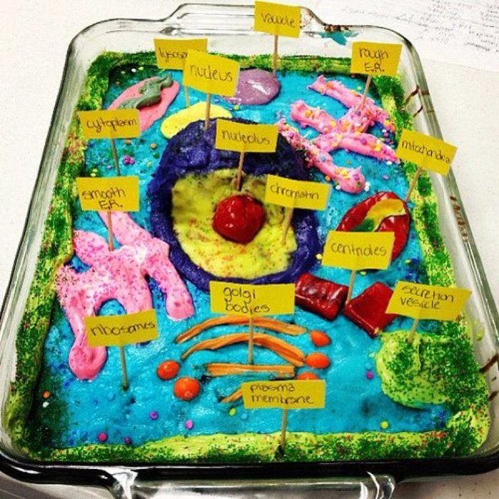
Fundamental Components of a Cell, 3d model of a cell project
Cells are composed of various components, each with a specific role in maintaining the cell’s life processes. These components are collectively known as organelles, and they work together in a coordinated manner.
- Cell membrane:The outer boundary of the cell, regulating the passage of substances in and out.
- Cytoplasm:The gel-like substance that fills the cell, containing various organelles and providing a medium for biochemical reactions.
- Nucleus:The control center of the cell, containing the genetic material (DNA) and regulating cellular activities.
- Ribosomes:Sites of protein synthesis, translating genetic information into functional proteins.
- Mitochondria:The powerhouses of the cell, responsible for generating energy through cellular respiration.
- Endoplasmic reticulum (ER):A network of interconnected membranes involved in protein synthesis, folding, and transport.
- Golgi apparatus:Processes and packages proteins and lipids for secretion or delivery to other organelles.
- Lysosomes:Cellular garbage disposals, breaking down waste materials and cellular debris.
Types of Cells
Cells exhibit a wide range of diversity in structure and function, adapting to specialized roles within multicellular organisms. These variations in cell types are crucial for the proper functioning of complex organisms.
- Prokaryotic cells:Simple cells lacking a nucleus and other membrane-bound organelles, found in bacteria and archaea.
- Eukaryotic cells:More complex cells with a nucleus and other membrane-bound organelles, found in plants, animals, fungi, and protists.
- Animal cells:Characterized by the presence of lysosomes and centrioles, which play important roles in cellular digestion and cell division.
- Plant cells:Distinctive for their rigid cell walls, chloroplasts for photosynthesis, and large vacuoles for storage and support.
History of Cell Research
The discovery and understanding of cells have been a long and fascinating journey, driven by the curiosity and ingenuity of scientists throughout history.
- Robert Hooke (1665):First observed and named “cells” while examining thin slices of cork under a microscope.
- Anton van Leeuwenhoek (1674):Developed improved microscopes and observed various single-celled organisms, including bacteria.
- Matthias Schleiden and Theodor Schwann (1838-1839):Proposed the cell theory, stating that all living organisms are composed of cells.
- Rudolf Virchow (1855):Added to the cell theory, stating that all cells arise from pre-existing cells.
3D Modeling Techniques
The creation of realistic and informative 3D cell models relies on a variety of techniques and software tools. Each technique offers unique advantages and disadvantages, depending on the specific needs of the model.
Software Options
Several software programs are commonly used for 3D modeling, each with its own strengths and limitations. Choosing the right software depends on the desired level of detail, complexity, and specific features required for the model.
- Blender:Open-source, versatile software with a wide range of tools for modeling, animation, and rendering.
- Maya:Industry-standard software for professional 3D animation, modeling, and visual effects.
- 3ds Max:Powerful software for architectural visualization, game development, and 3D modeling.
- ZBrush:Specialized software for digital sculpting and high-resolution modeling.
Modeling Techniques
Different modeling techniques are employed to create 3D models, each with its own strengths and weaknesses. The choice of technique depends on the desired level of detail, complexity, and efficiency.
- Polygon modeling:A traditional technique that involves creating models by manipulating polygons, providing control over shape and detail.
- NURBS modeling:Uses mathematical curves and surfaces to create smooth and organic shapes, suitable for modeling complex structures.
- Voxel modeling:Builds models by manipulating 3D pixels (voxels), offering a simplified and intuitive approach for creating blocky or stylized models.
Advantages and Disadvantages
Each modeling technique offers specific advantages and disadvantages in the context of cell representation.
| Technique | Advantages | Disadvantages |
|---|---|---|
| Polygon modeling | High level of detail, control over shape and topology | Can be time-consuming for complex models, requires knowledge of polygon geometry |
| NURBS modeling | Smooth and organic shapes, suitable for complex structures | Can be difficult to control for beginners, requires understanding of mathematical curves |
| Voxel modeling | Simplified and intuitive approach, efficient for blocky or stylized models | Limited detail, less control over shape and topology |
Designing the 3D Cell Model: 3d Model Of A Cell Project
Before embarking on the actual modeling process, it is essential to carefully plan the design of the 3D cell model. This planning phase ensures that the model accurately reflects the cell’s structure and function while maintaining visual appeal and clarity.
Key Features and Structures
The 3D cell model should include the key features and structures that are essential for understanding the cell’s function. These structures should be accurately represented in terms of their size, shape, and spatial relationships.
- Nucleus:The central control center of the cell, containing the DNA and regulating cellular activities.
- Cytoplasm:The gel-like substance that fills the cell, providing a medium for biochemical reactions.
- Mitochondria:The powerhouses of the cell, responsible for generating energy through cellular respiration.
- Ribosomes:Sites of protein synthesis, translating genetic information into functional proteins.
- Endoplasmic reticulum (ER):A network of interconnected membranes involved in protein synthesis, folding, and transport.
- Golgi apparatus:Processes and packages proteins and lipids for secretion or delivery to other organelles.
- Lysosomes:Cellular garbage disposals, breaking down waste materials and cellular debris.
Model Plan
A detailed plan Artikels the model’s dimensions, materials, and visual representation. This plan serves as a blueprint for the modeling process, ensuring consistency and accuracy.
- Dimensions:The model’s size should be scaled appropriately to represent the actual size of the cell and its components.
- Materials:The choice of materials should reflect the real-world properties of the cell’s structures, such as the membrane’s flexibility or the nucleus’s dense structure.
- Visual representation:The model should be visually appealing and informative, using colors, textures, and lighting to enhance clarity and understanding.
Hierarchical Structure
Organizing the model’s elements into a hierarchical structure allows for efficient editing and animation. This structure simplifies the process of selecting, manipulating, and animating individual components or groups of components.
- Top-level:The entire cell model.
- Sub-levels:Individual organelles, such as the nucleus, mitochondria, and ER.
- Lower levels:Components within organelles, such as the nuclear envelope or the mitochondrial cristae.
Creating the Model’s Components
Once the design plan is established, the next step is to create the individual components of the cell model. Each component should be meticulously modeled to accurately represent its shape, structure, and function.
Designing and Modeling
The modeling process involves using the chosen software and techniques to create each component’s geometry and shape. The level of detail should be appropriate for the intended use of the model.
- Nucleus:A spherical structure with a distinct nuclear envelope and nucleolus.
- Cytoplasm:A gel-like substance represented as a smooth surface with varying textures.
- Mitochondria:Bean-shaped organelles with folded inner membranes (cristae).
- Ribosomes:Small, spherical structures found in the cytoplasm or attached to the ER.
- Endoplasmic reticulum (ER):A network of interconnected membranes, either smooth or rough, depending on the presence of ribosomes.
- Golgi apparatus:Stacked, flattened sacs involved in processing and packaging proteins and lipids.
- Lysosomes:Small, spherical organelles containing enzymes for cellular digestion.
Textures and Materials
Appropriate textures and materials are used to create a realistic and informative representation of the cell’s components. These materials should reflect the real-world properties of the cell’s structures.
- Cell membrane:A thin, flexible membrane with a smooth or slightly textured surface.
- Nucleus:A dense, spherical structure with a smooth or slightly textured surface.
- Mitochondria:A smooth, bean-shaped structure with a folded inner membrane.
- Ribosomes:Small, spherical structures with a slightly rough surface.
- Endoplasmic reticulum (ER):A smooth or rough network of membranes, depending on the presence of ribosomes.
- Golgi apparatus:Stacked, flattened sacs with a smooth or slightly textured surface.
- Lysosomes:Small, spherical organelles with a smooth or slightly textured surface.
Characteristics and Functions
Each component of the cell model should be accompanied by information about its specific characteristics and functions. This information can be presented through text labels, annotations, or interactive elements.
- Nucleus:Contains the cell’s genetic material (DNA) and regulates cellular activities.
- Cytoplasm:Provides a medium for biochemical reactions and supports the cell’s organelles.
- Mitochondria:Generates energy through cellular respiration, providing the cell with ATP.
- Ribosomes:Synthesize proteins, translating genetic information into functional proteins.
- Endoplasmic reticulum (ER):Involved in protein synthesis, folding, and transport.
- Golgi apparatus:Processes and packages proteins and lipids for secretion or delivery to other organelles.
- Lysosomes:Break down waste materials and cellular debris, maintaining cellular health.
Assembling the 3D Cell Model
Once all the individual components have been created, they are assembled into a complete 3D cell model. This assembly process involves ensuring that the model accurately reflects the spatial relationships and interactions between cell structures.
Combining Components
The individual components are carefully positioned and aligned within the model to reflect their actual spatial relationships within a real cell. This process requires attention to detail and knowledge of cell biology.
- Nucleus:Positioned at the center of the cell, surrounded by the cytoplasm.
- Mitochondria:Scattered throughout the cytoplasm, often near the cell’s energy-consuming processes.
- Ribosomes:Found either free in the cytoplasm or attached to the ER.
- Endoplasmic reticulum (ER):A network of membranes that extends throughout the cytoplasm, connecting with the nuclear envelope.
- Golgi apparatus:Located near the ER, receiving proteins and lipids for processing and packaging.
- Lysosomes:Scattered throughout the cytoplasm, breaking down waste materials and cellular debris.
Spatial Relationships and Interactions
The model should accurately represent the spatial relationships and interactions between cell structures. This includes showing how organelles are connected, how they move within the cell, and how they interact with each other.
- Nucleus-cytoplasm interaction:The nucleus regulates cellular activities by sending messenger molecules (RNA) to the cytoplasm.
- ER-Golgi apparatus interaction:Proteins synthesized on the ER are transported to the Golgi apparatus for further processing and packaging.
- Mitochondria-cytoplasm interaction:Mitochondria generate ATP, which is used by the cell’s various processes, including protein synthesis and movement.
Optimization for Rendering and Animation
The assembled model should be optimized for efficient rendering and animation. This involves simplifying the model’s geometry, reducing the number of polygons, and optimizing the materials and textures.
- Geometry optimization:Reducing the number of polygons while maintaining the model’s visual fidelity.
- Material optimization:Choosing materials and textures that are efficient for rendering.
- Animation optimization:Simplifying the model’s animation to reduce rendering time and maintain smooth movement.
Enhancing the Model’s Visual Appeal
A visually engaging and informative 3D cell model can greatly enhance understanding and engagement. Various techniques can be employed to enhance the model’s visual appeal, making it more captivating and memorable.
Lighting and Shading
Appropriate lighting and shading effects can create a sense of depth, realism, and visual interest. This involves using different light sources, adjusting shadows, and experimenting with materials to create a visually appealing and informative model.
- Ambient lighting:Provides a general illumination, creating a sense of atmosphere and depth.
- Directional lighting:Simulates the sun or other distant light sources, casting shadows and highlighting features.
- Point lighting:Creates a focused light source, simulating a lamp or other nearby light source.
Animation
Animation can be used to demonstrate cell processes and functions, bringing the model to life and making it more interactive. This involves creating keyframes and using motion paths to animate the movement of organelles and other cell components.
- Protein synthesis:Animating the movement of ribosomes along the ER and the translation of mRNA into protein.
- Mitochondrial respiration:Animating the movement of molecules through the mitochondrial membrane and the production of ATP.
- Cell division:Animating the process of mitosis, showing the separation of chromosomes and the formation of two daughter cells.
Visualization Techniques

Various visualization techniques can enhance the model’s impact and understanding. These techniques can include using different color schemes, highlighting specific features, and adding interactive elements.
- Color coding:Using different colors to represent different organelles or cell structures.
- Transparency:Making certain components transparent to reveal internal structures.
- Interactive elements:Adding clickable elements that provide additional information or animations.
Final Wrap-Up
By building a 3D model of a cell, we unlock a deeper understanding of the microscopic world that surrounds us. This project empowers us to visualize complex biological processes, fostering a deeper appreciation for the intricacies of life itself.
Whether used for educational purposes, scientific research, or medical advancements, this project offers a powerful tool for exploring the wonders of the cellular realm.
Top FAQs
What software is best for creating a 3D cell model?
The best software depends on your experience and the complexity of your model. Popular options include Blender, Maya, and ZBrush. Each software has its own strengths and weaknesses, so it’s worth exploring them to find the best fit for your project.
What are some common mistakes to avoid when designing a 3D cell model?
Avoid oversimplifying the cell structure. While a basic model is a good starting point, it’s crucial to include as much detail as possible to accurately represent the cell’s complexity. Also, be mindful of scale and proportion to ensure the model is accurate and visually appealing.
How can I make my 3D cell model more engaging?
Experiment with lighting and shading effects to enhance visual appeal. Consider using animation to showcase cell processes and functions. You can also explore interactive features that allow viewers to explore the model in 3D space.
